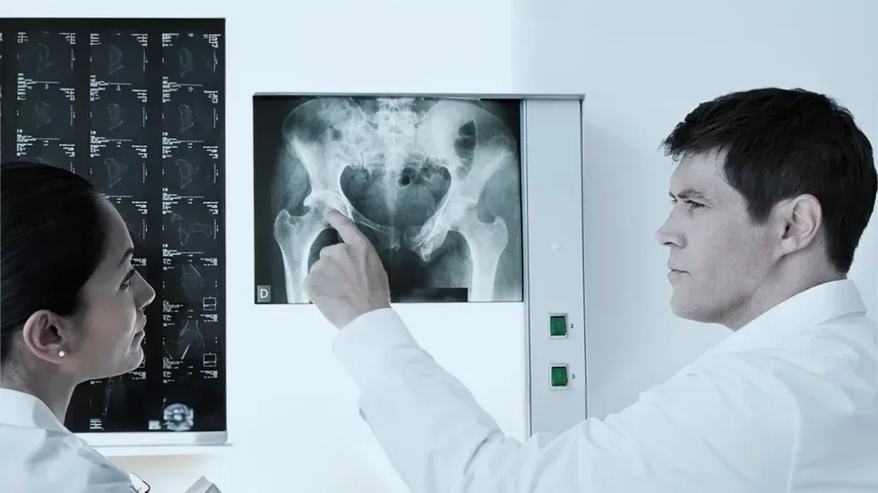
Many patients present with unclear complaints in the groin and hip region. Often numerous doctor consultations take place beforehand.
With appropriate experience, diseases can be reliably diagnosed using standard diagnostic procedures (examination by a specialist, X-ray, ultrasound, MRI).
In addition to the radiological findings, we also evaluate X-ray images and MRI ourselves. In special cases, an arthro-MRI (contrast medium) or a 3D MRI motion analysis is carried out.
If a bone correction of the hip is necessary, this can be simulated with this system before the operation. In this way, we can guarantee the best possible surgical result.
Your orthopedist in Berlin will be happy to help you effectively and competently.
Why Choose Our Diagnostic Approach?
At Sportorthopädie Berlin, we combine decades of experience with state-of-the-art diagnostic technology. Dr. Alexander Moser and his team take the time to understand your individual symptoms and develop a personalized diagnostic plan.
The combination of thorough clinical examination, advanced imaging, and when indicated, diagnostic injections, allows us to achieve diagnostic precision that many patients have not experienced elsewhere.
Dr. Alexander Moser
Your hip specialist in Berlin
”Hip surgery at the highest level”
Hip surgery experts Berlin – Hip pain Berlin – Hip joint pain Berlin – Groin pain Berlin
Watch the explainer video
Frequently asked questions
What diagnostic steps are commonly taken for groin and hip pain?
<p>– <strong>Medical History:</strong> The first step to a correct diagnosis is always a targeted medical history. The doctor records the patient's medical history and asks about specific symptoms. Specific physical stresses, for example at work or during sporting activities, can also be relevant.</p><p>– <strong>Clinical Examination:</strong> The physical examination is of crucial importance. It identifies movement restrictions, tenderness and possible trigger factors. The range of motion (ROM), specific functional tests (Impingement Test, Apprehension Test, Subspine Test, FEAR Test, Leg roll Test, etc.) and pain localization allow conclusions to be drawn about anatomical causes of hip pain.</p><p>– <strong>Imaging Procedures:</strong> X-rays, MRI or CT to visualize structural changes in the groin or hip area. X-rays represent spatial forms better than cross-sectional imaging (CT and MRI). Today, often only an MRI of the hip is performed. However, many conditions (such as acetabular dysplasia) are difficult to assess in MRI and are often overlooked. Standard X-rays (pelvic overview, Lauenstein view, Dunn view, false profile view) are often necessary for an exact diagnosis. For surgical planning, the X-rays can be calibrated digitally, allowing surgical planning (e.g., prosthesis planning or planning of bony corrections for femoroacetabular impingement) to be carried out on the computer before the operation. This increases the precision of the hip surgery.</p><p>– <strong>Modern 3D Diagnostics:</strong> Modern diagnostics already allow three-dimensional representations and motion analyses (4D motion analyses) using MRI or CT imaging. If necessary, such imaging is initiated.</p><p>– <strong>Ultrasound Examination:</strong> Ultrasound of the hip plays an important role in the diagnosis of groin and hip pain:</p><p>• Visualization of soft tissues: Ultrasound enables detailed visualization of soft tissue structures such as muscles, tendons, ligaments and bursae, which is particularly relevant for identifying injuries or inflammation.</p><p>• Inguinal hernias: Ultrasound is an effective method for diagnosing inguinal hernias, as it allows dynamic visualization during certain movements.</p><p>• Motion analysis: Dynamic movement tests to assess the functionality of the hip and identify any movement restrictions or irregularities.</p><p>• Guidance for interventions: Ultrasound can also be used during certain therapeutic interventions, for example for targeted injection of medication into the hip region.</p><p>• Risk-free method: Compared to other imaging procedures, ultrasound is non-invasive and without radiation exposure, making it a safe option.</p><p>– <strong>Laboratory Tests:</strong> Blood tests can reveal inflammatory processes or other systemic causes.</p><p>– <strong>Diagnostic Joint Infiltration (Joint Injection):</strong> Joint infiltration of the hip refers to the injection of medication, usually anti-inflammatory corticosteroids and a local anesthetic, directly into the hip joint. This injection can have therapeutic and diagnostic purposes:</p><p>• Therapeutic aspect: Infiltration of medication into the hip joint can help reduce inflammation and relieve pain. This is particularly useful for conditions such as arthritis or bursitis.</p><p>• Diagnostic aspect: Joint infiltration also serves as a diagnostic tool. If the pain decreases significantly after the injection, this may indicate that the hip joint is the source of pain. This confirms or rules out the hip as the primary cause of the complaints.</p><p>• Motion analysis: Infiltration can also be performed in connection with diagnostic movement tests to check whether the injection relieves or improves pain during certain movements.</p><p>The combination of these examinations enables a more accurate diagnosis and lays the foundation for developing a suitable treatment plan.</p>
What is the importance of medical history in the examination of groin and hip pain?
<p>The medical history in the examination of groin and hip pain includes important steps:</p><p>– <strong>Pain characteristics:</strong> The doctor asks about the type of pain (stabbing, dull, pulling, etc.) to differentiate possible causes.</p><p>– <strong>Onset and duration of symptoms:</strong> When did the pain first appear and how has its intensity changed over time?</p><p>– <strong>Trigger factors:</strong> Are there certain movements, activities or loads that intensify or trigger the pain?</p><p>– <strong>Previous illnesses and injuries:</strong> Information about previous injuries, operations or chronic diseases can provide clues to possible causes.</p><p>– <strong>Activity level:</strong> The doctor asks about the patient's usual activity level to determine whether certain movements or sports influence the complaints.</p><p>– <strong>Accompanying symptoms:</strong> The presence of accompanying symptoms such as swelling, numbness or movement restrictions is determined.</p><p>A thorough medical history is crucial to obtain a comprehensive picture of the complaints and serves as a starting point for further diagnostic measures.</p>
What role does clinical examination play in the diagnosis of hip pain?
<p>– <strong>Determine movement restrictions:</strong> Checking the mobility of the hip joint provides information about possible problems such as arthritis or impingement.</p><p>– <strong>Check tenderness:</strong> The doctor examines the area around the hip for tenderness to identify possible inflammation or injuries.</p><p>– <strong>Assess gait pattern:</strong> A precise assessment of walking can indicate certain hip problems or muscular imbalances.</p><p>– <strong>Perform provocation tests:</strong> Specific tests help localize the cause of pain, e.g., through rotational movements or flexions of the hip joint. Special function tests include: Impingement Test, Apprehension Test, Subspine Test, FEAR Test, Leg roll Test, etc.</p><p>The clinical examination serves as a starting point for further diagnostic steps, including imaging procedures such as X-rays, MRI or CT, to enable a precise diagnosis.</p>
What imaging techniques are used to diagnose groin and hip problems?
<p>– <strong>X-rays:</strong> Enable visualization of bone structures and can show bone problems such as fractures or osteoarthritis.</p><p>– <strong>Magnetic Resonance Imaging (MRI):</strong> Provides detailed images of muscles, ligaments, tendons and soft tissues to identify injuries or inflammation.</p><p>– <strong>Computed Tomography (CT):</strong> Used for detailed imaging of bones and is particularly suitable for assessing bony structures.</p><p>– <strong>Ultrasound:</strong> Used to examine soft tissue structures, especially if inguinal hernias are suspected.</p><p>These procedures enable an accurate diagnosis by visualizing the structural aspects of groin and hip problems. Depending on the clinical suspicion, the doctor decides which imaging procedure is most suitable.</p>
How do you differentiate between groin and hip pain in diagnostics?
<p>The differentiation between groin and hip pain is carried out through a thorough clinical examination and, if necessary, additional specific tests:</p><p>– <strong>Medical history:</strong> The doctor asks about the medical history and checks whether certain movements or activities trigger the pain.</p><p>– <strong>Pain localization:</strong> Through precise localization, an attempt is made to determine whether the pain originates from the hip or the groin area.</p><p>– <strong>Mobility tests:</strong> Examinations that check the mobility of the hip and groin help identify the pain-causing region.</p><p>– <strong>Imaging procedures:</strong> X-rays, MRI or CT visualize structural changes in the hip and groin and help with differentiation.</p><p>– <strong>Ultrasound:</strong> Particularly if an inguinal hernia is suspected, ultrasound can be a targeted examination method.</p><p>– <strong>Joint injection (joint infiltration):</strong> If pain decreases significantly after targeted injection into the joint under ultrasound guidance, this indicates that the hip joint is the source of pain. This diagnostic tool has very high value when a reliable diagnosis was not possible after standard procedures.</p><p>The combination of these approaches enables a precise diagnosis and differentiation between groin and hip pain.</p>
Which laboratory tests can be helpful in diagnosing groin and hip problems?
<p>Laboratory tests can help identify systemic causes or inflammatory processes. Here are some relevant tests:</p><p>– <strong>Blood count:</strong> Can indicate signs of infections or inflammation.</p><p>– <strong>Inflammation markers (CRP, ESR):</strong> Increased values can indicate inflammatory processes.</p><p>– <strong>Rheumatoid factors and antibodies:</strong> Tests to rule out autoimmune causes.</p><p>– <strong>Electrolytes and kidney function tests:</strong> Particularly in long-lasting or severe complaints, to rule out systemic problems.</p><p>– <strong>Blood sugar tests:</strong> Diabetes can affect the joints and lead to complaints.</p><p>Not every patient requires comprehensive laboratory tests. Whether specific tests are necessary depends on the individual medical history and clinical findings.</p>
What specific tests are done to diagnose inguinal hernias?
<p>The diagnosis of inguinal hernias can be supported by various specific tests:</p><p>– <strong>Cough or Valsalva test:</strong> The patient is asked to cough or strain strongly while the doctor examines the groin area. This helps identify a hernia, as the pressure during coughing can intensify the protrusion.</p><p>– <strong>Inguinal canal palpation:</strong> The doctor palpates the groin area to look for swelling or a palpable protrusion.</p><p>– <strong>Ultrasound examination:</strong> Ultrasound enables dynamic imaging of the groin area and is effective in identifying hernias.</p><p>– <strong>CT or MRI:</strong> These imaging procedures can be used to more accurately localize and assess the size of an inguinal hernia.</p><p>If, after our diagnostics, there is suspicion of an inguinal hernia, we can recommend one of the leading specialists for inguinal hernia treatment upon request.</p>
How is the differential diagnosis made between muscular, bony and nerve-related groin and hip pain?
<p>The differential diagnosis requires a comprehensive clinical assessment and can include various diagnostic approaches:</p><p>– <strong>Medical history:</strong> Detailed recording of the medical history, including the type and duration of pain, previous injuries and accompanying symptoms.</p><p>– <strong>Clinical examination:</strong> The doctor checks mobility, tenderness and muscle function in the groin and hip area.</p><p>– <strong>Imaging procedures:</strong> X-rays, MRI or CT can show structural changes in bones, joints and soft tissues.</p><p>– <strong>Electromyography (EMG) and nerve conduction velocity:</strong> These tests can reveal nerve damage or compression.</p><p>– <strong>Blood tests:</strong> Can indicate inflammatory processes or systemic causes.</p><p>– <strong>Functional tests:</strong> Special tests to assess muscle function, biomechanical processes and nerve function.</p><p>– <strong>Joint infiltration:</strong> Targeted injection under ultrasound guidance enables differentiation of pain causes. In unclear cases, it has a very high diagnostic value.</p><p>The combination of these methods makes it possible to differentiate between muscular, bony and nerve-related causes.</p>